 Corporate Website
 Africa
 Argentina
 Asia
Asia
 Australia
 Belgium
 Brazil
 Bulgaria
 Canada (EN)
 Chile
Chile
 China
China
 Colombia
 Denmark
 Egypt
Egypt
 France
 Germany
 Greece
 Hungary
 Indonesia
 Italia
 India
India
 Japan
Japan
 Korea
Korea
 Malaysia
 Mexico
 Middle East
 Netherlands
 Peru
Peru
 Philippines
 Poland
 Portugal
 Romania
 Russia
 South Africa
 Spain
Spain
 Sweden
 Thailand
 Tunisia
 Turkey
 Ukraine
 United Kingdom
 USA
USA
 Vietnam
The Ceva Lung Program offers the methodology and guidelines on how to correctly evaluate the presence, incidence, circulation patterns and impact of Mycoplasma hyopneumoniae, Actinobacillus pleuropneumoniae and Aujeszky's disease virus infections using serological investigation and adapted lung scoring of slaughter pigs.
It is used to determine the appropriate vaccination protocol and monitor the results of vaccination. This brochure will bring insight into slaughter house lung checks in pigs.
1. Mycoplasma hyopneumoniae
Mycoplasma hyopneumoniae (M. hyo) is the primary pathogen causing Enzootic Pneumonia (EP), one of the most prevalent respiratory diseases in pigs resulting in high morbidity but low mortality. It is also one of the main pathogens involved in the porcine respiratory disease complex (PRDC), however other bacteria and viruses are also often contributing factors.
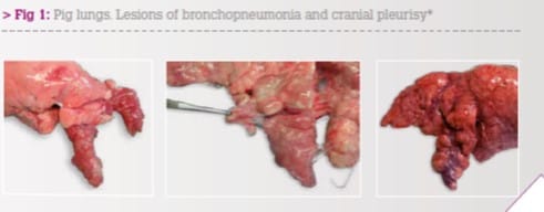
Acute inflammation, due to M. hyo infection, leads to a catarrhal or even purulent exudate in the airways. The accumulation of neutrophils and mono-nuclear cells around the bronchi and bronchioles results in airway collapse and consolidation of the lungs.
Lung tissue infected with M. hyo develops consolidation and catarrhal broncho-pneumonia with purple to grey regions of meaty appearance. The consolidation can be observed from 3 - 12 weeks post infection. The lesions are mainly localised in the apical and cardiac lobes, in the anterior part of the diaphragmatic lobes, as well as in the intermediate lobe. Lesions resolve after 12 to 14 weeks with formation of interlobular fissures, also referred to as scarring.
M. hyo and related viral and bacterial pathogens are also responsible for the development of pleuritis, which is typically localised in the cranio-ventral part of the lungs, most often between the apical and cardiac lobes.
Consolidation of the apical and cardiac lung lobes indicating the acute to sub-acute bronchopneumonia is suggestive of M. hyopneumonia induced EP. Pleuritis at the junction of the cardiac and diaphragmatic lobes can still be assigned to the cranio-ventral area. Pleurisy in this area are most likely due to M. hyo and associated bacterial or viral infections.
2. Actinobacillus pleuropneumoniae
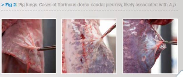
The forms of pleurisy located in the dorso-caudal region are primarily caused by Actinobacillus pleuropneumoniae (A.p.) and to a lesser extent by other pathogens.
A.p. is responsible for serious acute clinical disease characterised from a pathoanatomical standpoint by cases of fibrino-necrotic-haemorrhagic pleuropneumonia, with variable mortality in animals afflicted according to the virulence of the serotype involved.
The chronic form of the infection is characterised by the development of chronic lung lesions appearing as fibrous pleurisy of variable gravity at the dorso-caudal side of lungs that can seriously impair the animal's productive career. This is because the dorsal pleural lesions, even more than the apical lesions, impose a mechanical limitation and a level of pain during the animals' respiratory movements that result in reduced daily weight gain and an increased number of days necessary for reaching slaughter weight.
The chronic pleural lesions, still present at the time of slaughter, can be easily observed in this context and provide key information regarding the health conditions of the farm, the timeliness of undertaking control measures or for assessing the results obtained by implementing them.
3. Evaluation of pneumonia and pleurisy
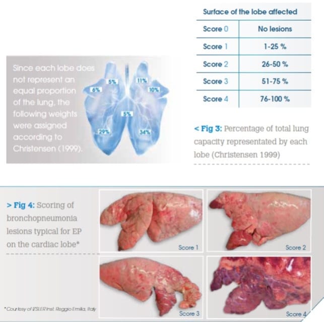
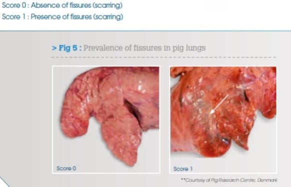
Pneumonia and pleurisy can be evaluated in a qualitative and quantitative manner. In the Ceva Lung Program, pneumonia is scored based on Madec method, which has been modified considering the contribution of each lobe to the overall capacity of the lungs. Quantification of Scarring is included. Pleurisy is scored using the Slaughterhouse Pleurisy Evaluation System (SPES) method, although here in the Ceva Lung Program all cranial pleurisy is recorded as Cranial Pleurisy Scoring. Then the APPI is calculated for the A.p. induced dorso-caudal pleurisy.
Modified Madec Scoring
For the modified Madec score, the enzootic pneumonia-like lung lesions of each lobe are quantified according to the following:
Quantification of Scarring
The prevalence of fissures, or scarring, is evaluated based on their presence or absence. The incidence of fissures indicates the old infections likely due to M. hyopneumoniae.
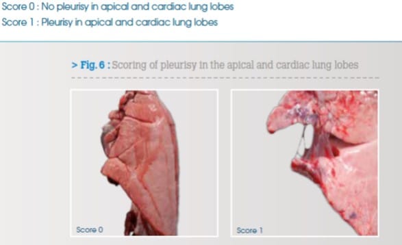
Cranial Pleurisy Scoring
Cranial pleurisy can be attributed to pathological processes in this area which are likely related to M. hyo and other respiratory pathogens such as SIV, Pasteurella multocida and other bacteria. This cranial pleurisy should be recorded separately from the dorso-caudal pleurisy to allow the appropriate differential diagnosis. These lesions include pleurisy on the surface of the lung lobes or between lobes as interlobar pleurisy. Pleurisy in the apical and cardiac lung lobes are scored as follows:
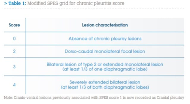
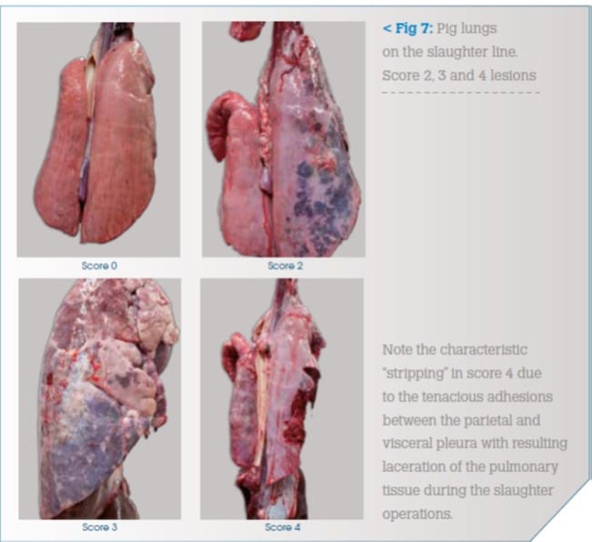
Dorsocaudal pleurisy based on SPES
The SPES method facilitates the assessment of pleural lesions according to their location, appearance and extension. The SPES method is based on a point system from 0 to 4 based on the presence, the extension and position of pleurisy observed on both lungs of each animal directly on the slaughter line (table 1).
- Processing the Scores
Through statistical processing of recorded data, the Ceva Lung Program enables not only measurement of the incidence of the injury, but also identification of the distribution by classes within the sample and restoration of the data in easily legible graphic displays. The Ceva Lung Program is capable of providing information about the frequency, severity and suspected origin of pathological changes most likely due to M. hyo and A.p.
This information includes:
- Enzootic pneumonia like lesions
1. Average percent of affected surface out of all lungs:
Using the Madec score assigned to each lobe, after taking into account the weight of that lung lobe in comparison to the entire lung, we are able to determine the percentage of the total lung surface affected for each animal. These values are then summed and divided by the total number of lungs scored to determine the average percent of affected surface out of all lungs evaluated. This gives the actual value of the lungs that are damaged by Enzootic pneumonia on average in the group of evaluated animals.
Average percent of affected surface out of all lungs = Sum of individual % of affected lung surface / N° of all scored lungs.
2. Average percent of affected surface out of pneumonic lungs:
This gives us the information about the extent of the lesions and thus severity of the infection in the sick animals. It is possible that a severe infection in a small number of animals can be diluted due to a large number of healthy animals within the group. This situation will be revealed with this calculation.
Average percent of affected surface out of lungs with active pneumonia = Sum of individual % of affected lung surface / N° of sick lungs
3. Average percent of scarring in pneumonic lungs:
This parameter reveals the percentage of resolved lesions attributed to Enzootic pneumonia. This indicates old infections likely due to M. hyo in the herd.
Average percent of scarring out of all lungs = N° of lungs with scars / N° of all scored lungs * 100
- Cranial pleurisy
Cranial pleurisy is scored based on its presence or absence. The percent of cranial pleurisy in the evaluated group indicates the probability of the previous infection of pathogens affecting the cranial part of the lungs, especially M. hyo. This value completes the evaluation of the lung, as we are able to quantify the presence of pathogens that did not result in long term parenchymal lesions.
Percent of cranial pleurisy = N° of lungs with cranial pleurisy / N° of all scored lungs * 100
- Actinobacillus Pleuropneumoniae Index (APPI)
The Actinobacillus pleuropneumoniae Index (APPI) provides information regarding the prevalence and seriousness of the dorso-caudal pleurisy, highly indicative of prior pleuropneumonia due to A.p.
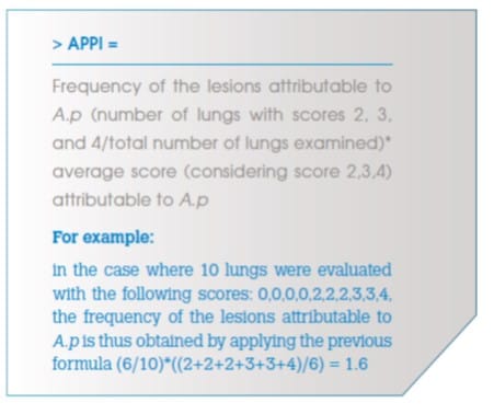
According to the original SPES method, a high level of correlation was found between the A.p. seropositivity of herds and the SPES value. This confirms the specificity of recording of the dorso-caudal pleurisy for previous A.p. infections.
4. Conclusion
The Ceva Lung Program Scoring Methodology is designed to assist in identifying the correct diagnosis of respiratory disease through the evaluation of lungs at slaughter. It enables the discovery of even subclinical infections that were not noted during the growing period.
The calculate values allow the estimation of both the incidence and the severity of enzootic pneumonia and pleuropneumonia. It can also be used to evaluate the efficacy of the control measures against M. hyo and A.p. when used both prior to and after their implementation.
Finally, when used routinely, the Ceva Lung Program can aid in the recognition of the dynamics of those infections in a defined period or seasonal differences.